CONGESTIVE HEART FAILURE
DEFINITION
It is the abnormal clinical condition involving impaired cardiac pumping that results in pathophysiologic changes of vasoconstriction & fluid retention leading to further increase in cardiac workload & eventually heart fails to contract adequately & maintain effective cardiac output
CAUSES
- Coronary artery disease: Coronary Arteries supply blood to the heart, when the coronary arteries are obstructed by a plaque or lesion there is inadequate blood supply to the myocardium, where the Myocardium is deprived of oxygen & fails to contract and eject enough blood into the Aorta
- Myocardial infarction: Due to sudden changes in the blood flow to myocardium can result in scarring of the Myocardial cells, that cannot contract & eject the blood and maintain cardiac Output
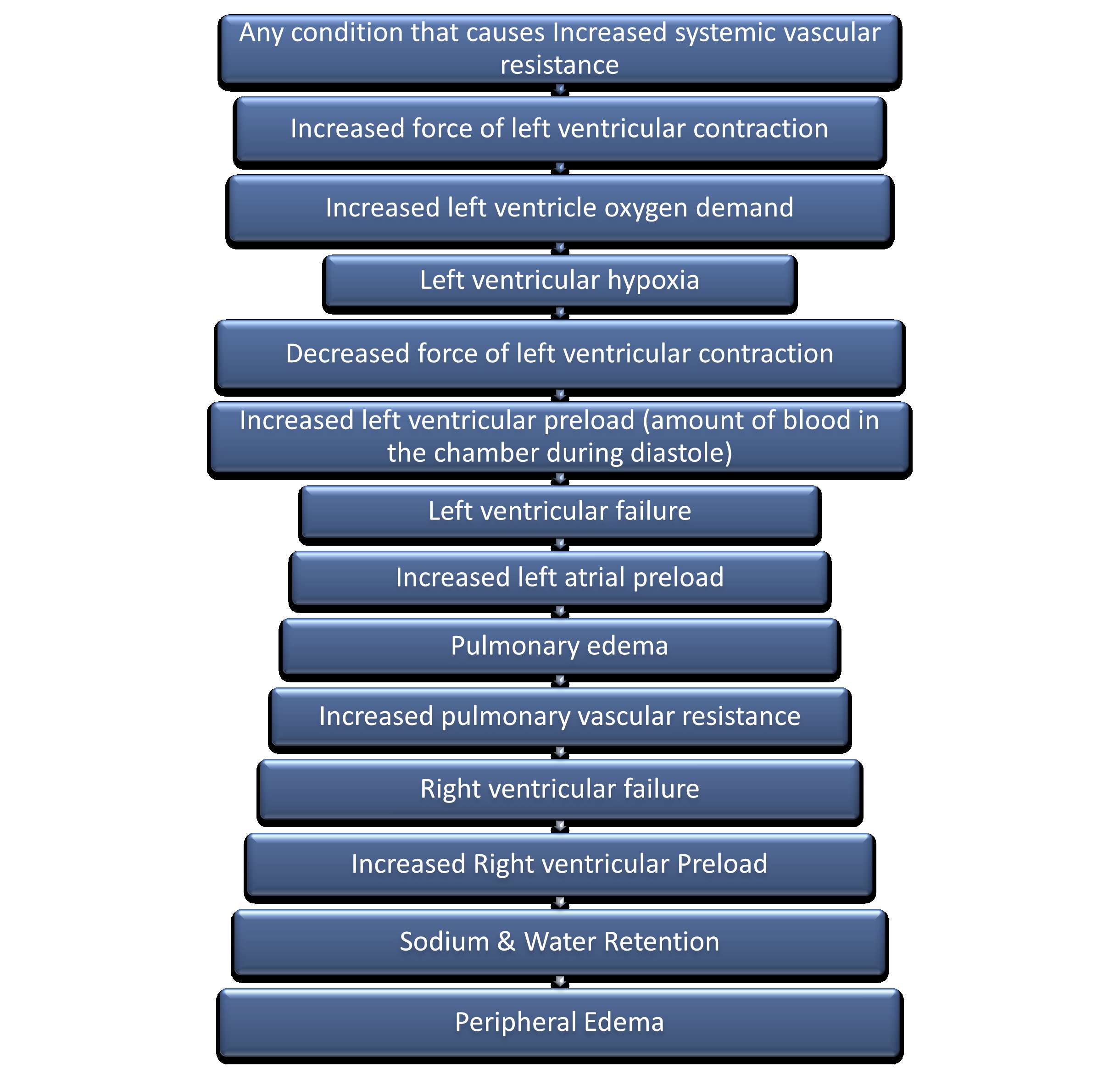
- Hypertension: Hypertension causes increased systemic vascular resistance which increases the force of Myocardial contraction & increased Myocardial oxygen demands, which eventually leads to failure of the heart to contract effectively and move the blood forward. As a result there is backflow of blood & congestion leading to heart failure
- Dysrhythmias
- Myocardial disorders like Cardiomyopathy & Myocarditis: Myocardial disorders can result impaired Myocardial contractility & failure to move the blood forward and maintain cardiac output
- Congenital heart disease: These are birth defects, characterized by ventricular dysfunction & heart failure
- Cor pulmonale (pulmonary heart disease): It is the condition characterized by right ventricular hypertrophy & right heart failure due to obstruction of blood flow in the lungs primarily caused by Chronic Obstructive Pulmonary Disease (COPD)
- Valvular heart disorders: These disorders can lead to valvular stenosis or regurgitation, where the valves are unable to eject the blood forward completely as a result backflow of blood & congestion occurs that leads to ventricular dilation & impaired Myocardial contraction
- Anemia: Due to decreased oxygen carrying capacity of the blood the Myocardium lacks oxygen supply which eventually result in decreased Myocardial contractility & ventricular dysfunction
- Inflammatory diseases of the heart like Rheumatic heart disease, Infective endocarditis: Inflammatory diseases of the heart can lead to valvular dysfunction, where valves fails to move the blood forward, as a result backflow of blood & congestion takes place, which makes the wall of the ventricles dilated & become resistance to systole (ventricular contraction)
- pulmonary emboli: It is the moving blood clot obstructing the pulmonary arteries, that results in lack of blood oxygenation in the lungs and backflow of blood in to the right ventricles leading to right ventricular hypertrophy & right sided heart failure
- Thyrotoxicosis: It is a thyroid disorder that causes altered metabolism of myocardial tissue as a result the Myocardium is deprived of oxygen and fails to contract effectively & maintain cardiac output
- Other Disorders: like Diabetes Mellitus & Kidney disorders can also cause congestive heart failure due to fluid retention & electrolyte Imbalances
CLASSIFICATION OF HEART FAILURE
- Systolic failure: It is the defect in which there is inability of the ventricles to pump causing increased ventricular filling and dilatation. the left ventricle loses its ability to generate enough pressure to eject blood forward through the aorta. Eventually the left ventricle becomes thin walled, dilated & hypertrophied that results in decreased Ejection Fraction (It is the percentage of total ventricular filling volume that is ejected during each ventricular contraction
- Diastolic failure: It is the impaired ability of the ventricles to relax & fill during diastole due to stiff and noncompliant ventricles. Decreased filling of the ventricles will result in decreased Stroke volume & cardiac Output. Diastolic Failure is characterized by high filling pressures & results in venous engorgement in both of the pulmonary & systemic vascular system
- Mixed systolic & diastolic failure: It is seen in cases of cardiomyopathy that is characterized by poor systolic function that is compromised by ventricular hypertrophy and ventricles inability to relax. Here the myocardial muscle fibers are weakened & results in poor systolic function, and is further compromised by dilated left ventricular walls that fails to relax.
- Left sided heart failure: this occurs due to left ventricular dysfunction which prevents normal flow of blood and causes blood to backflow in to the left atrium & pulmonary vasculature. The increased Pulmonary pressure causes fluid extravasation from the pulmonary capillary bed into the interstitium then into the alveoli causing pulmonary congestion & edema
- Right sided heart failure: This occurs due to increased backflow of blood in to the pulmonary arteries and right ventricle it leads right ventricular congestion & failure, resulting in venous congestion & peripheral Edema
PATHOPHYSIOLOGY OF HEART FAILURE
https://learnnursingeasy.co.in/storage/uploads/ck/nursing175100171231.jpg
CLINICAL MANIFESTATION
Right sided failure:
- Right ventricle heaves- visible pulsation on the chest wall
- cardiac murmurs
- Jugular venous distension
- Edema
- Weight gain
- Increased heart rate
- Ascites
- Anasarca (whole body edema)
- Hepatomegaly
- Anxiety & depression
- Rt. Quadrant Abdominal pain
- Anorexia & GI distension
- Nausea & vomiting
Left sided failure:
- Left ventricle heaves
- pulsus Alternans (alternate strong and weak pulse felt on palpation)
- Respiratory crackles due to pulmonary edema
- Abnormal heart murmurs
- Altered mental status like restlessness, confusion & anxiety
- Depression
- Dyspnea
- Shallow respirations
- Paroxysmal nocturnal dyspnea
- Orthopnea
- Dry cough and frothy pink sputum
Associated Symptoms
- Weakness
- Fatigue
- Nocturia
- Decreased Exercise Tolerance
- Altered Sensorium
- Wheezing
- Heart Palpitations/ Irregular Heart beat
DIAGNOSTIC STUDIES
- History & physical examination to assess the type of failure
- Cardiac enzymes like CPK (>200mg/dl) and CK-MB (>20mg/dl) are elevated
- Complete Blood Count determines Anemia with Increased ESR Count
- ABG analysis determines Low Oxygen Saturation with Acid base Imbalances
- Liver function test shows Elevated Liver enzymes
- Sr. Electrolytes determines electrolyte Imbalances with altered Renal Function
- Bleeding & Clotting Profile are dearranged
- Chest X-ray to detect cardiomegaly and Rt. or Lt. axis deviation
- ECG denotes Right or Left Ventricular Hypertrophy with ST segment Changes
- Echo Cardiography shows Valvular Dysfunction with changes in Chamber dimensions
- Sr. B-type natriuretic peptide (>100pg/ml) is elevated
- Nuclear imaging studies like MUGA Scans are done to monitor for Ventricular hypertrophy and Increased Cardiac Volume
- Cardiac catheterization to identify the cardiac output & detect the chamber pressures.
MANAGEMENT
- Treat the underlying cause: Correction of MI through Coronary Artery Bypass Grafting (CABG) surgery, Hypertension with antihypertensive drugs, Cardiomyopathy with heart transplantation
- Improving Gas Exchange & Oxygenation: Oxygen therapy with mask or Non-invasive ventilatory support or Bilevel positive airway pressure or with Endotracheal Intubation & Mechanical ventilation is provided as required
- Decrease Intra vascular volume: Inj. Loop diuretics like inj. Lasix to decrease fluid overload & decrease the venous return to the ventricles thus reducing the preload and allowing the overfilled ventricles to contract effectively & maintain cardiac output
- Hemodialysis with ultra filtration: This procedure allows the removal of excess intravascular fluid through a peripheral or central venous catheter without significantly changing the mean arterial pressure
- Decreasing Venous Return: This is achieved by placing the client in high fowler's position, as a result blood flows into the extremities
- Increasing Myocardial Oxygen Supply: Inj. Nitroglycerine is a vasodilator administered to reduce preload & increase Myocardial Oxygen supply
- Decreasing Preload(ventricular stretching) & Afterload (resistance against which the left ventricle has to pump): Inj. IV nitroprusside is a vasodilator that decreases preload & afterload, improves Myocardial contraction, increases Cardiac Output & relieves pulmonary congestion. Inj. Morphine Sulphate, also reduces preload & afterload, treats chest pain & pulmonary Edema
- Vasoactive Therapy: Inj. Nesiritide is a recombinant form of B-type natriuretic peptide causes both Arterial & venous dilation
- Improving Cardiac Function: Ionotropic Agents like Inj. Digoxin to increase ventricular contractility; ß-adrenergic Agonists like Dopamine, Dobutamine, Epinephrine & Nor-epinephrine is administered to increase Myocardial contractility
- Treating Dysrhythmias: inj. Cordarone or Amiodarone is administered to treat cardiac dysrhythmias. Defibrillation to revert severe dysrhythmias.
- Reducing Anxiety: Sedatives like Benzodiazepines & Anxiolytics like Alprazolam is administered to relieve Anxiety
INVASIVE THERAPIES
- Intra aortic Balloon Pump: It is a sausage shaped balloon that is inserted through femoral access and advanced into the aorta which is connected to a pneumatic helium device which inflates and deflates alternatively with heart’s pumping action which is done temporarily to support the heart by increasing ventricular contractility.
- Ventricular assist devices:
- A ventricular assist device (VAD) is also known as a mechanical circulatory support device that helps the heart to pump blood from the chambers (ventricles) to the all parts of the body. VAD is implanted by performing an open heart Surgery.
- It is done as a temporary procedure before a heart transplantation until a donor heart is available or permanently to help the heart maintain circulation.
- Ventricular assist devices can be implanted in one or both ventricles of the heart, but it is most frequently implanted in the left ventricle & called as left ventricular assist device (LVAD).
- Cardiac Resynchronization Therapy(CRT):
- CRT is done to restore the normal coordinated pumping action of the ventricles by overcoming the delay in electrical conduction caused by the left bundle branch block, which is an event of heart failure
- In this procedure, a specialized pacemaker called a CRT device or Bi-Ventricular pacemaker is implanted through the vein and is accessed to the right atrium along with the standard wires (leads) that are implanted in the right atrium and right ventricle present in a normal dual chamber pacemaker, a third wire (lead) is implanted to stimulate the lateral wall of the left ventricle. The lateral wall of the left ventricle is paced via the coronary sinus which is a vein that wraps around the back of the heart, around the left ventricle.
- Once the LV lead is positioned, the CRT device will be programmed to pace the right ventricle and left ventricle simultaneously. This leads to improved co-ordination of the heart’s pump function and can be seen as a narrowing of the QRS in the ECG.
- This device sometimes may also contain an implantable cardioverter-defibrillator (ICD), which can deliver an electrical shock to revert the heart's normal Sinus rhythm, in case of serious dysrhythmias.
SURGICAL MANAGEMENT
- Heart transplantation: this is the treatment of choice for patients with severe heart failure.
It is of 2 types
- Orthotopic transplantation: In which the recipient heart is removed and the donor’s heart is transplanted.
- Heterotopic transplantation: In which the recipient heart is left in place and the donor’s heart is placed near anastomosing it with the right atrium.


