
Loading...


Structure of Bone
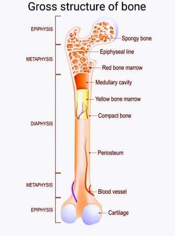
The Anatomic Structure of bone is best studied using a typical long bone. Each long bone consists of the Epiphysis, the diaphysis and the metaphysis.
LAYERS OF THE BONE:
Bone consists of two layers called the Periosteum and Endosteum
GROSS STRUCTURE OF FLAT BONES:
Flat bones vary extensively in their surface features depending on the principal function and location in the body. These bones are expanded into broad flat plates that provide surfaces for muscle attachment. The flat bones in the body are the bones of the cranium or skull (Occipital, Parietal, frontal, Nasal, Lacrimal &vomer; the coxal or hip bones (Ilium, Ischium & Pubis) and the sternum, ribs and scapula
Flat bones are composed of two thin layers of compact bone (lined on either side) enclosed in between a variable quantity of cancellous bone called as diploe, which is the location of red bone marrow. In cranial bones, the layers of compact tissue are usually termed as tables of the skull, the outer layer is thick and tough and the inner layer is thin and brittle, hence termed as vitreous (glass like tables)
MICROSCOPIC STRUCTURE OF BONE
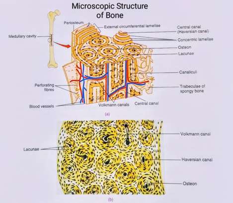
Bone contains both Organic material (Collagen) and inorganic material (Calcium & phosphate). The internal and external growth and the remodeling of the bone are ongoing process. Based on the structure bones are classified as cortical (Compact and dense) and cancellous (Spongy bone)
Compact bone is stronger & denser bone tissue that is found interior to the periosteum of the long bones. It is composed of cylindrically shaped structural units that fit closely together in the compact bone, creating a dense bone structure called the Osteon or haversian systems. At thecentre of each osteon, central canals run parallel to the bone’s long axis called as the haversian canals that contains blood vessels, nerves and lymphatic vessels. These vessels and nerves branch off at right angles through a canal, called the volkmann’s canal that travels to the bones interior from the periosteum and extends to the endosteum. The haversian canals are surrounded by concentric rings of calcified matrix called the lamellae, which comprises the mature bone. Smaller canals, called canaliculi extend from the haversian canals to the lacunae, where the mature bones are embedded.
Cancellous bones contain osteocytes surrounded by lacunae, but they lack the organized structure because the lamellae are not arranged in concentric circles as in cortical or compact bone. Instead the lacunae and osteocytes are found like a network of bone matrix spikes called as trabeculae, which are filled with red or yellow bone marrow. Each trabecula forms along the line of stress to provide strength to the bone. The spaces in the trabecular network provide balance by distributing the weight to the dense and heavy compact bone by making the bone lighter and facilitate easy movement of the muscles.