INTRODUCTION
The Axial Skeleton is made up of 80 bones that form the central core of the body. It consists of skull, hyoid bone, vertebral column, the ribs and the sternum. The Axial Skeleton protects the major vital organs of the body like the brain, Spinal cord, heart and lungs.
⦁ Skull: the Skull consists of the 8 cranial bones and 14 facial bones along with the 6 middle ear ossicles or Auditory bones
⦁ Cranium: The cranial bones also called the Neurocranium that forms the upper part of the skull and it protects the brain and brain stem. The cranial bones are as follows
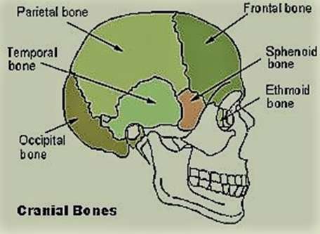
⦁ 2 Parietal bones (Right and left)
⦁ 2 Temporal Bones ( Right and left)
⦁ 1 Frontal Bone
⦁ 1 Occipital bone
⦁ 1 Ethmoid Bone
⦁ 1 Sphenoid Bone
- Parietal Bones: The two large parietal bones articulate in the centre to form the sagittal suture. These are cranial bones that form the side and top (roof) of the head. Anteriorly the two parietal bones Join with the frontal bone, posteriorly is the Occipital bone and below are the temporal and Sphenoidal bones. The Squamous suture seperates the parietal and temporal bones.
- Temporal Bones: The two temporal bones are situated at the base and sides on each side of the skull lateral to the temporal lobes of the cerebrum.
- The temporal lobe consists of four regions namely; the squamous, mastoid, petrous and tympanic regions. The squamous is the largest and most superior region; Inferior to the squamous is the mastoid region; in between the two fused region is the petrous region and the small tympanic region lies inferior and anterior to the mastoid.
- Temporal lobe consists of two processes namely, Zygomatic process and the Styloid Process. The Zygomatic process projects from the lower squamous region and articulates with the Zygomatic bone of the cheek; the styloid process projects downwards from the interior of the temporal lobe and provides attachment to the muscles of the tongue.
- The temporal lobe has four borders namely, the occipitomastoid suture (Seperates the occipital bone and mastoid region); the squamousal suture (seperates the parietal bone and squamous region); the sphenosquamosal suture (seperates the sphenoid bone and squamous region); the zygomaticotempral suture (Seperates the zygomatic bone and zygomatic process of the temporal bone)
- Frontal Bone: The frontal bone is an unpaired bone that forms the anterior and superior portions of the skull.
- The frontal bone Consists of three regions namely; squamous region (which is the large flat portion) that forms the main region of the fore head; Orbital region (which lies inferiorly) and forms the superior region of the orbit; nasal region (which is a small part) that articulates with the nasal bones and maxilla to form the roof of the nose
- The frontal bone has 2 borders namely; the coronal suture (where it attaches to the parietal bone) and the sphenofrontal suture (where it attaches to the Sphenoid bone)
- The frontal bone also articulates with the zygomatic, nasal bones and the maxilla
- Occipital Bone: the Occipital bone forms the base of the skull at the back of the cranium. It contains the foramen magnum, the large opening of the skull through which the spinal cord passes and enters into the vertebral column. It articulates with the first vertebra of the Spinal cord
- The Occipital bone has two borders namely; lambdoidal suture (where it borders with the parietal bone) and the occipitomastoid suture (where it borders with the temporal bone)
- Ethmoid Bone: The ethmoid bone is a small bone in the skull that separates the nasal cavity from the brain. It is a spongy light weight bone with air-filled construction and is located at the roof of the nose between the two orbits. The Ethmoid bone is centrally located and in the Neurocranium it articulates with the frontal and Sphenoid bone. It also articulates with several bones in the viscerocranium.
- Sphenoid Bone: The Sphenoid bone is a butterfly shaped bone with wing like processes located in the middle of the skull towards the front and forms the back of the orbit.
- Sphenoid bone consists of parts namely body of the bone, 2 greater wings, 2 lesser wings and the pterygoid processes. The body of the bone forms the middle part and articulates with the ethmoid and occipital bone and forms the key region of the nasal cavity, it also contains the sphenoid sinuses. The greater wings form the floor of the middle cranial fossa and the lesser wings projects laterally and form the floor of the anterior cranial fossa and the superior orbital fissure through which there is passage for the optic nerve.
Facial Bones: The facial bones are also called as the viscerocranium, they support the soft tissues of the face and provides shape and characteristic features for the face. The facial bones are joined together with fibrous immovable Joints that attaches to the Skull, except the Mandible which is attached to the skull by a movable tempero-mandibular Joint that facilitates the movement of the lower Jaw for mastication and Speech. The facial skeleton provides protection for the brain and also serves as passage for Neuro vascular Structures. The facial bones are as follows.
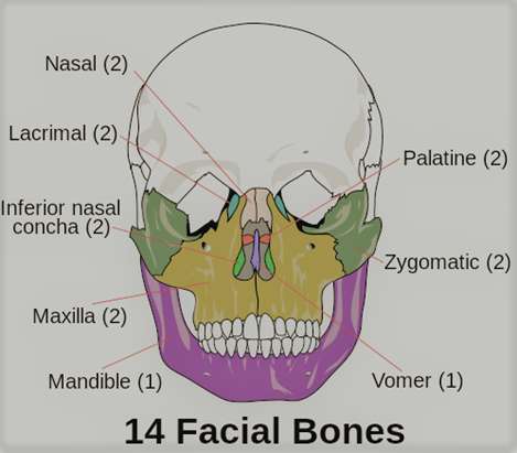
2 maxilla Bones (Right and left)
⦁ 2 Zygomatic Bones (Right and left)
⦁ 2 Nasal Bones (Right and left)
⦁ 2 Inferior Nasal Concha (Right and left)
⦁ 2 Palatine Bones (Right and left)
⦁ 2 Lacrimal Bones (Right and Left)
⦁ 1 Mandible
⦁ 1 Vomer
- Maxilla: The two maxillary bones forms the lining for the upper teeth, the floor for the nose and the base for the orbits. The maxilla fuses in the midline and form the upper Jaw. They articulate with the Zygomatic, nasal, lacrimal and palatine bones.
- Zygomatic Bones: The two Zygomatic bones form the cheeks and contribute to the orbits, they articulate with the frontal, temporal, maxilla and Sphenoid bones
- Nasal Bones: The two nasal bones are slender and located in the midline of the face. The two bones fuse to form the bridge of the nose. They articulate with the frontal, ethmoid and maxillary bones
- Inferior Nasal Concha: The two inferior Nasal conchae bones are located within the nasal cavity. They are made up of spongy bones and have a curly shape. These bones increase the surface area of the Nasal cavity thus increasing the amount of air that contacts the mucus membrane and cilia of the nose. Their primary function is to filter or purify, warming and humidification of the air before entering the lungs.
- Palatine Bones: The two palatine bones fuse in the midline to form the palatine. These bones are located at the back of the nasal cavity and form the roof for the mouth and floor for the orbits.
- Lacrimal Bones: The two lacrimal bones are the smallest bones in the face. These two bones form the medial wall of the orbit and articulate with the frontal, ethmoid, maxilla, Inerior Nasal conchae.
- Mandible Bone: The mandible forms the movable lower part (jaw) of the skull. The upper parts of the facial bones are attached to the cranium and the lower part forms the mandible. The Joint between the mandible and the temporal bones of the cranium is called temoromandibular Joint, that forms the only non-sutured mobile joint in the skull.
- Vomer Bone: At the base of the nasal cavity is the small vomer bone that forms the nasal Septum
Middle ear Ossicles: There are six (6) bones located in the middle ear (3 in each ear) in the tympanic cavity. They are responsible to transit the sound waves from the tympanic membrane to the inner ear and aid in hearing.
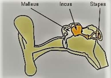
2 Malleus (Right and left ear)
⦁ 2 Incus (Right and left ear)
⦁ 2 stapes (Right and left ear)
- Malleus: Malleus is the small hammer shaped first ossicle or bone located in the middle ear. It consists of head, neck, short process and handle. The handle of malleus is loosely attached to the inner surface of the Tympanic Membrane and moves to transfer the sound waves. The head of the malleus is attached to the incus with a incudo-malleolar joint.
- Incus: The incus is the second Ossicle that attaches with the head of the malleus. It contains a body, short and long processes. The incus connects with the Stapes by a incudo-stapedial joint and serves to transfer the sound waves
- Stapes: The Stapes is the third stirrup shaped middle ear ossicle that consists of head, body and foot plate. It is the smallest bone of the human body. The foot plate of the stapes attaches to the oval window and transfers the sound waves to the inner ear.
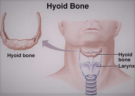
The hyoid Bone: the hyoid bone is a horse shoe shaped bone also called as lingual or tongue bone located below the lower jaw in the anterior midline of the neck between the chin and thyroid cartilage and is attached by the muscles to the tongue and larynx. It is the only bone that is not articulated with any other bone nearby but distantly articulated to bones with muscles and ligaments.
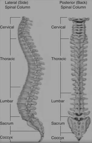
The Vertebral Column: The vertebral column is also called as backbone or spine that consists of 33 vertebrae that includes 7 cervical, 12 thoracic, 5 lumbar, 5 sacral and 4 coccygeal vertebrae. The upper 24 pre-sacral vertebrae are separated each other by Intervertebral discs and the 5 sacral and 4 coccygeal vertebrae are fused bone in adults. The vertebral column is characterized by several curved structure with vertebral body, arch and processes. A typical vertebra varies in size, shape and characteristics especially they differ from one region to another and no two vertebrae are identical. The vertebral column provides attachments to muscles, supports the trunk, protects the spinal cord and nerve roots and serves as a site for haemotopoiesis.
VERTEBRAL COLUMN
- Cervical Vertebrae (C1-C7): The cervical spine begins from the end of the Occipital bone present at back of the head. The cervical vertebrae are further divided in to two as: Upper cervical vertebrae (C1 & C2) and the Lower cervical vertebrae (C3 to C7). The first cervical vertebra (C1) is called the Atlas vertebra that supports the skull and its appearance differs from other vertebrae by a ring of bone made up of two lateral masses joined at the front and back by the anterior arch and the posterior arch. The second Cervical vertebra is called the Axis (C2) with a blunt tooth like process that projects upwards in the form of dens that allows movement of the head as the atlas rotate around the dens.
- Thoracic Vertebrae (T1-T12): The thoracic vertebral body is large compared to the cervical vertebra. The typical characteristic feature of thoracic vertebra is that it contains a spinous process, which is a long protruded downward angle that allows attachment to the next inferior vertebra. The superior articular processes of thoracic vertebra face anteriorly and the inferior processes face posteriorly. The thoracic vertebra also contains several additional articulation sites, which are referred to as facets which provide articulation with the head end of the ribs.
- Lumbar Vertebrae (L1- L5): The lumbar vertebrae are characterized by large size and thickness of the vertebral body as they bear the greater weight of the body. They have short transverse processes and blunt spinous processes that project posteriorly. The articular process are large, with the superior processes facing backward and inferior processes facing forward.
- Sacral Vertebrae: The sacrum is a triangular bone with the thick and wide superior base, where it is major weight bearing, and then tapers at the inferior apex, where it is non-weight bearing. It is formed by 5 fused sacral vertebrae, on the anterior surface, the vertebral lines of fusion are seen as 4 transverse ridges and on the posterior surface along the midline is a bumpy ridge that is the remnant of the fused spinous process referred as median sacral crest. Similarly the transverse processes of the sacral vertebra forms the lateral sacral crest.
- Coccyx: the Coccyx, also referred as a tail bone is forms by the fusion of 4 very small coccygeal vertebrae. It articulates with the inferior tip of the sacrum.
The ribs: The ribs are the long curved bones and are 24 in number; there are 12 pairs of ribs on each side attached to the sternum in the center that forms the bony framework of the thoracic cavity and protects the thoracic organs. Each rib articulates posteriorly with two thoracic vertebrae by the costovertebral Joint, except for the first rib, as it attaches only to the thoracic vertebrae.
The ribs are classified into three groups as True, false and floating ribs based on their attachment with the sternum.
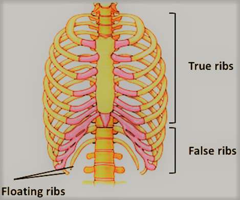
- True ribs: The true ribs are directly attached to the sternum with their costal cartilage. They articulate to the sternum with Sternocostal Joints, except the first rib that uniquely articulates with the clavicle by costoclavicular Joint.
- False ribs: False ribs are the 8th, 9th, 10th ribs that indirectly articulate with the sternum as their costal cartilage connects with the 7th Costal cartilage by the costochondral Joint.
- Floating ribs: The 11th & 12th ribs are the distal ribs that do not articulate with the sternum at all and thus referred to as floating ribs.
The typical ribs consist of head, neck and body or shaft. The Head is wedge shaped with two articular facets separated by a wedge of the bone. One facet articulates numerically corresponding to the thoracic vertebrae and other facet articulates with the above vertebrae. The neck simply connects the head with the body through a rough tubercle and articulates with the transverse process of the corresponding vertebrae. The Body or shaft of the rib is flat and curved. The internal surface of the shaft has a groove that provides neurovascular supply to the ribs and provides protection to the nerves and blood vessels.
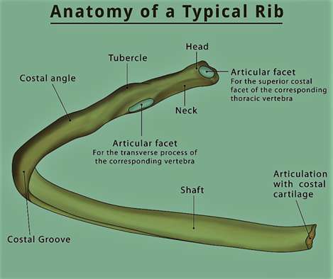
The atypical ribs are the 1st, 2nd, 10th, 11th & 12th ribs that differ from their features that are common to all ribs
⦁ The first rib is atypical because it is wide and short, has two costal grooves, and one articular facet.
⦁ The second rib is thin, long, and has a tuberosity on its superior surface for the attachment of the serratus anterior muscle.
⦁ The tenth rib has only one articular facet.
⦁ The eleventh and twelfth ribs have only one articular facet with no neck.