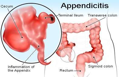
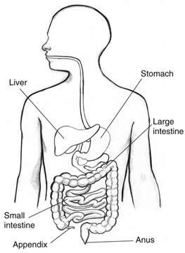
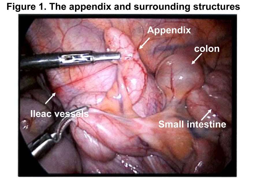
Anatomy
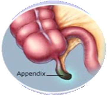
- The appendix is vestigial organ present in the right lower abdomen attached to the cecum which is a fingerlike pouch that measures about 10cm (4 inches).
- The inside of the appendix forms a cul-de-sac that opens into the large intestine.
- The inside of the appendix is called the appendiceal lumen.
- Mucus created by the appendix travels through the appendiceal lumen and empties into the large intestine.
- When that opening gets blocked, the appendix swells and can easily get infected by bacteria.
Definition
- test
- Appendicitis is a condition characterized by inflammation of the appendix that causes painful swelling in the right lower abdomen, as the inflammation worsens the pain becomes severe and requires immediate medical attention or surgery to remove the infected appendix.
Causes
Sources of obstruction include
- faeces, parasites, or growths that clog the appendiceal lumen
- Enlarged lymph tissue in the wall of the appendix causing compression and blocks the appendiceal lumen
- Inflammatory bowel disease (Crohn’s disease and ulcerative colitis )
- Kinking of appendix
- Trauma to the abdomen
- Fibrous condition of bowel wall
- External occlusion by adhesions
- Bacterial infection in wall of appendix
Risk factors
- Anyone at any age can get appendicitis
- More common among people 10 to 30 years old.
- 7% of the population
- Very young and old because they have a higher rate of complications.
Pathophysiology
Primary obstruction of the appendix lumen

Appendix filled with mucus and swells

Increase pressures in the lumen and walls of appendix

Occlusion of blood vessels, reduced venous drainage

Result in thrombosis and occlusion of small vessels

Stasis of lymphatic flow
Ischemia & Necrosis of Appendix

Bacteria leak out through the dying walls

Pus forms within and around the appendix (suppuration)

Appendiceal rupture

Peritonitis
Symptoms
- Pain
- The abdominal pain occurs before other symptoms
- Gets worse in a matter of hours
- Gets worse when moving around, taking deep breaths, coughing, or sneezing
- Localized findings in the right iliac fossa.
- The abdominal wall becomes very sensitive to palpation.
- Rebound tenderness
Pain Signs
-
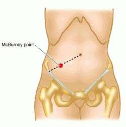
- McBurney's sign
- Rovsing sign
- Psoas sign
- Blumberg sign
McBurney point
- McBurney point a point of special tenderness in appendicitis, about one-third the distance between the right anterior superior iliac spine and the umbilicus.
Rovsing's sign
- Continuous deep palpation starting from the left iliac fossa may cause pain in the right iliac fossa, by pushing bowel contents and thus increasing pressure around the appendix.
Psoas Sign
- This is right lower-quadrant pain that is produced with the patient lying on his left side and then extending the hip. Because extension elicits pain, the patient will lie with the right hip flexed for pain relief.
Blumberg sign
- Palpation of the left iliac fossa, followed by sudden release causes contralateral (right iliac fossa) rebound tenderness.
Other symptoms of appendicitis may include
- loss of appetite
- Nausea
- Vomiting
- Constipation or diarrhea
- Inability to pass gas
- a low-grade fever that follows other symptoms
- abdominal swelling
- Feeling that passing stool will relieve discomfort.
Complications
- Perforation: Ulceration and necrosis of the Appendix
- Peritonitis: Inflammation of the peritoneum due to rupture of Appendix
- Abscess formation: Localized pus collection in the abdominal cavity.
- Septicemia: Infection of blood.
Diagnostic tests
- History & Physical examination confirms the diagnosis on palpation
- CBC – Elevated WBC counts due to Infection
- ESR raised due to infection
- Ultrasound shows the presence of diseased Appendix
- CT scan to confirm the diagnosis
- Alvarado scoring
|
Alvarado score for Acute Appendicitis
|
|
Feature
|
score
|
|
Symptoms
|
|
Migartion of pain
|
1
|
|
Anorexia
|
1
|
|
Nausea
|
1
|
|
Signs
|
|
Right Iliac fossa tenderness
|
2
|
|
Rebound tenderness
|
1
|
|
Fever
|
1
|
|
Laboratory
|
|
Leucocytosis
|
2
|
|
Shift to left (segmented Neutrophils
|
1
|
|
Total
|
10
|
|
Alvarado score for Acute Appendicitis
|
|
Score 1-4: Acute Appendicitis, very unlikely keep under observation
|
|
Score 5-6: Acute Appendicitis, may be for regular observation
|
|
Score 7-8: Acute Appendicitis. Probable operate
|
|
Score 9-10: Acute Appendicitis, definite, operate
|
Medical Management
- Pain management
- NPO status
- Ice cap application to reduce inflammation
- Prepare for surgery
- Antibiotics
- IV fluids
Surgical Management
- Open Appendectomy: It is performed as open surgery using one abdominal incision about 2 to 4 inches (5 to 10 centimeters) long. Open surgery is done, If the appendix has ruptured and infection has spread beyond the appendix or there is an abscess, where an open appendectomy is essential to clean the abdominal cavity.
- Laparoscopic appendectomy: Surgery is done through a few small abdominal incisions & by inserting a special surgical tools and a video camera through the Incision to remove the diseased appendix. In Laparoscopic surgery there is faster recovery and healing with less pain and scarring. It may be better for older adults and people with obesity.
Pre Operative Care
- NPO
- IV fluids
- Antibiotics such as ceftriaxone and metronidazole
- to help kill bacteria
- reduce the spread of infection in the abdomen
- Reduce postoperative complications
- If NPO surgery is done under General Anesthesia, Otherwise, spinal anesthesia may be used.
- Shave and prepare
- Antibiotics along with pain medication may also be administrated prior to appendectomies.
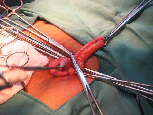
Open Appendectomy
- Patient is placed under general anesthesia
- The incision is two to three inches long and it is made in the right lower abdomen, several inches above the hip bone.
- Once the incision opens the abdomen cavity and the appendix is identified
- The surgeon removes the infected tissue and cuts the appendix from the surrounding tissue.
- Closing the incision using surgical staples or stitches to close the skin up.
- In order to prevent infections the incision is covered with a sterile bandage
Laparoscopic Appendectomy
- The newer method to treat appendicitis
- This surgical procedure consists of making three to four incisions in the abdomen, each 0.25 inches to 0.5 inches long.
- Inserting laparoscope into one of the incisions.
- The laparoscope is connected to a monitor to help surgeon to inspect infected area in the abdomen.
- The other two incisions are made for the specific removal of the appendix by using surgical instruments.
- Laparoscopic surgery also requires general anesthesia and it can last up to two hours
Post Operative Care
- The recovery process may vary depending on the severity of the condition
- Limit physical activity so the tissues can heal faster.
- Recovery after an appendectomy does not request diet changes or a lifestyle change.
- vital signs closely monitored
- Pain medication
- offer clear liquids the day after the surgery and then progress to a regular diet
- Patients sit up on the edge of the bed and walk short distances for several times a day.
- Full recovery from appendectomies takes about 4 to 6 weeks
Post Operative Complications
- Infection
- Bleeding
- Thrombophlebitis
- Pneumonia
Nursing Assessment
-
- Assess pain, location and type
- Asses for presence of complications
- Assess vital signs, Intake/ Output Chart
- Monitor Lab values as CBC, Electrolyte counts
- Monitor C-reactive Protein
Nursing Management
- Administer IV fluids as prescribed to prevent dehydration
- Antibiotic Therapy with Sensitive Antibiotics to treat Infection
- Analgesics like Diclofenac Sodium/ Tramazac to relieve Pain
- Reduce Anxiety for clients undergoing surgery by providing Psychological support
- Continuous monitoring of the Surgical clients
- Surgical dressing to the incised area following strict Aseptic Technique
- Meeting the Nutritional needs of the client with soft solids & bland diet
- Follow up Services & Education on home care Management.